Figure 2.
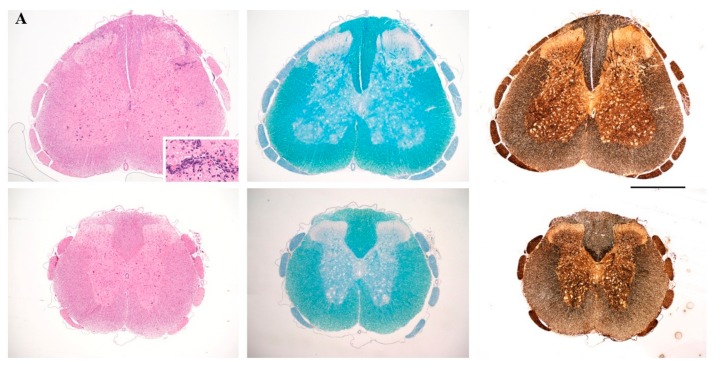
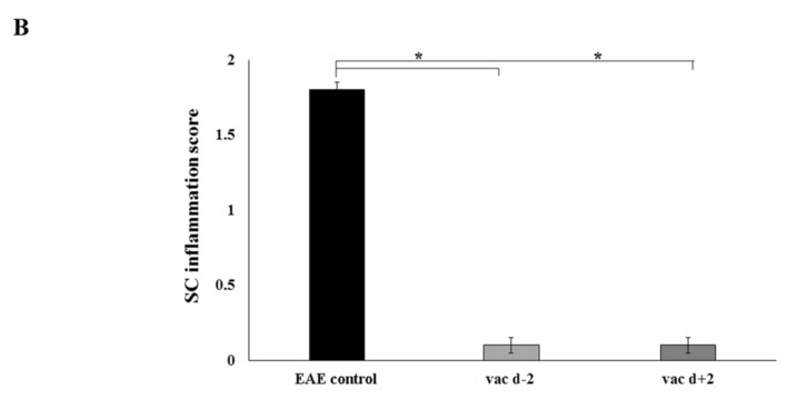
(A) Neuropathological analysis of spinal cord sections taken on day 17 following immunization, from representative nonvaccinated control rats (top) and rats vaccinated, two days before (prophylactic) induction of myelin basic protein 72–85 epitope (MBP72–85)-induced experimental autoimmune encephalomyelitis (EAE), with cyclo(87–99)[Arg91, Ala96]MBP87–99 (bottom). Sections were stained with H&E for inflammation (left panels), Luxol Fast Blue for myelin (central panels), and Bielschowsky’s silver stain for axons (right panels). Inset shows inflammatory infiltrates in the spinal cord parenchyma. Representative sections from 1 animal per group are shown (n = 3 for each group). Scale bar 1 mm; (B) Semi-quantitative representation of spinal cord (SC) inflammation in nonvaccinated EAE control rats (EAE control), rats vaccinated with cyclo(87–99)[Arg91, Ala96]MBP87–99, 2 days before (vac d − 2) and 2 days after (vac d + 2) induction of MBP72–85-EAE. Pathological changes of spinal cord sections were scored as inflammatory infiltrates/mm2 of tissue for the three groups of rats (n = 3 for each group). Statistical significance after multi-group comparisons, using the Mann–Whitney test, is shown (*, p < 0.05).


