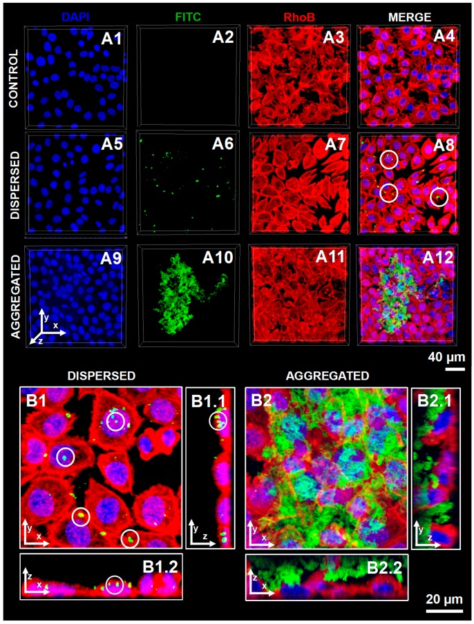
Figure 4.
CLSM images of L929 cells upon contacting with well-dispersed or aggregated (FITC-CHT/ALG)2 hollow multilayered microcapsules. (A1–A4) Cells that were not in contact with microcapsules were used as controls. Dispersed (FITC-CHT/ALG)2 hollow multilayered microcapsules were internalized by cells (A5–A8). Contrarily, aggregated capsules were not incorporated and remained adsorbed on the cell surface (A9–A12). A zoomed-in region of (A8,A12) micrographs was done to distinguish the internalized dispersed capsules (B1–B1.2) from the ones adsorbed at the cell surface due to their higher aggregation degree (B2–B2.2). (B1,B2) correspond to 3D reconstructed z-stack micrographs as seen from top (view of the xy plane). B1.1–B2.1 and B1.2–B2.2 correspond to orthogonal projection images (view of the yz plane and view of the xz plane, respectively). Blue channel: DAPI nuclear probe; Red channel: rhodamine-labeled phalloidin; Green channel: (FITC-CHT/ALG)2-based hollow multilayered microcapsules. White circles indicate some examples of microcapsules internalized by L929 cells.

