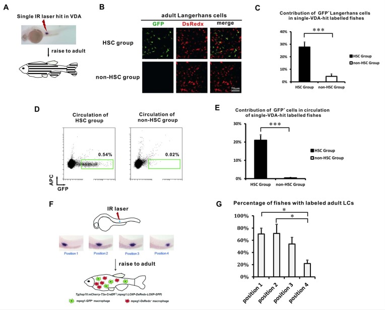
Figure 3. The VDA-derived LCs correlate with HSCs spatially.
(A) A schematic view of the single spot VDA heat-shocked experiment. A single IR laser hit is performed in the VDA region. The heat-shocked embryos are raised to adult (about 3 months) for analysis. (B) GFP+ LCs are mainly found in the HSC group but not in the non-HSC group of VDA labeling. (C) Quantification of the relative contribution of GFP+ LCs in HSC (n = 8) and non-HSC (n = 5) groups. Error bars represent mean SEM. ***p<0.001. (D) Flow cytometry shows GFP+ cells are mainly found in the circulation of HSC group but not in the circulation of non-HSC group. (E) Quantification of the relative contribution of GFP+ cells in the circulation of HSC (n = 8) and non-HSC (n = 5) groups. Error bars represent mean SEM. ***p<0.001. (F) A schematic view of the position restricted single spot VDA heat-shocked experiment. The VDA region from anterior to posterior is artificially divided into four positions (P1 to P4) and each position is labeled by a single IR laser hit. The embryos are raised to adult and the percentage of successful LCs labelling is analyzed for each labeled position. (G) The percentage of fish with widely labeled LCs in each position. Three independent experiments were performed. There are total 35 fish for position 1, 44 fish for position 2, 40 fish for position 3, and 38 fish for position 4. *p<0.05.

