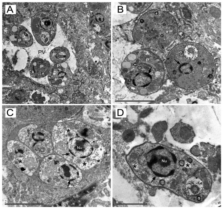
Figure 5.
Transmission electron microscopy of mice skin lesion. The ultrastructural analysis of amastigotes from lesion were performed in untreated (A) or mice treated with meglumine antimoniate or epoxymethoxylawsone at higher doses (22.7 mg of Sb(V)/kg/day and 11.4 mg/kg/day, respectively). In (A): amastigotes with no morphological changes within parasitophorous vacuoles (PV); bar-shaped kinetoplast (K); nuclei (N) and flagellar pocket (Fp). In (B): Amastigotes (P) show dense nuclear chromatin (thin arrow) with altered profile and absence of nucleoli (→). In (C): Amastigotes (P) with rarefied cytoplasm (*); kinetoplast with an atypical condensation (k) and dense nuclear chromatin (→) and nucleolus (Nu). In (D): Amastigote (P) shows dense nuclear chromatin with altered profile and absence of nucleoli. The images are representatives of ten selections of each group mice.

