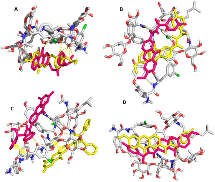
Figure 8.
Analyte 1 (A), Analyte 14 (B), Analyte 30 (C), and Analyte 18 (D), docked on teicoplanin selector. Chiral selectors are represented in sticks with C, O, N, and Cl atoms colored in grey, red, blue, and green, respectively. (S) and (R) enantiomers are represented with magenta and yellow sticks, respectively. Hydrogen interactions and π-stacking interactions are represented with dashes and double arrow, respectively.

