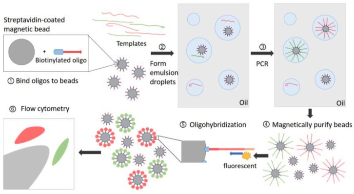
Figure 5.
BEAMing procedure. Step 1: Magnetic beads coated with streptavidin are bound to biotinylated oligonucleotides (oligos). Step 2: An aqueous mix containing all the necessary components of PCR, beads and template DNA are stirred together with an oildetergent mix to create microemulsions. Red and green lines represent two kinds of templates which may differ from each other by one or more nucleotides. The aqueous compartments (blue circles in grey oil layer) are fully diluted to ensure each of them contain an average of less than one template molecule and less than one bead. Step 3: These microemulsions are subjected to thermal cycles of conventional PCR. If a DNA template and a bead are present to be together in a single aqueous compartment, the bead-bound oligos act as a primer for amplification. Step 4: Breaking the emulsions and the beads are purified with a magnet. Step 5: Fluorescently labeled primers are hybridized to the amplified target DNA molecules, which renders the beads as specific signal after appropriate laser excitation. Step 6: Flow cytometry is used to count the beads with different fluorescence signals.

