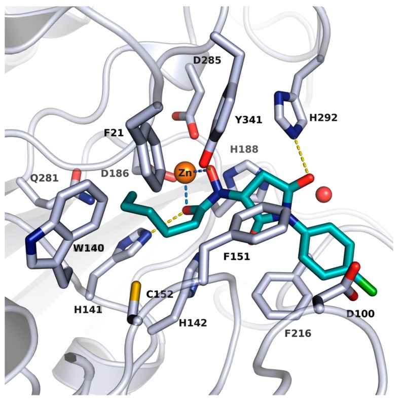
Figure 1.
Docking pose of J1036 in smHDAC8 (PDB ID: 4BZ8). Protein backbone is shown as a ribbon and side chains of key amino acid residues in the active site are shown as white sticks. The catalytic zinc ion and water molecule are shown as an orange and red sphere, respectively. Coordination of the zinc ion by J1036 is represented with light blue lines while the hydrogen bond interaction between the docked hit and the protein is shown as a yellow line.

