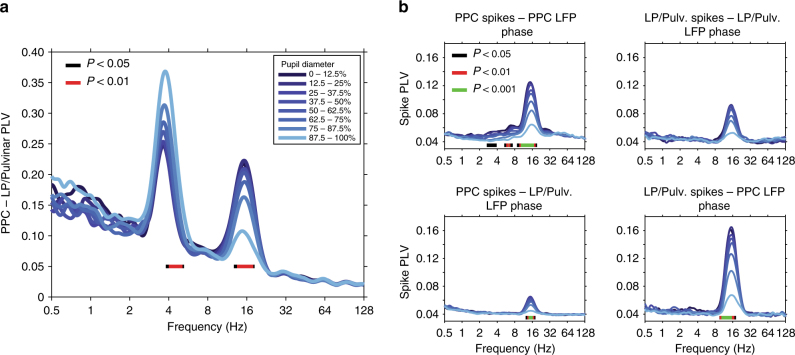
Fig. 3.
Thalamo-cortical synchronization varies with ongoing fluctuations in pupil-linked arousal. a PLV measured between LP/Pulvinar and PPC as a function of LFP frequency and pupil diameter. Pupil diameter is denoted by color (see legend). Note the prominent thalamo-cortical phase synchronization in the theta (∼4 Hz) and alpha (12–17 Hz) carrier frequency bands. Significant modulation across pupil diameter bins is indicated by bars plotted below PLV traces (one-way ANOVA, FDR-corrected P-values). PLV in the theta band significantly increased with pupil dilation, while alpha PLV significantly decreased with pupil dilation. b The phase synchronization (PLV) of spiking activity to LFP rhythms recorded within the same brain structure (top row), as well as between regions (bottom row). Spike PLV both locally within PPC and LP/Pulvinar, as well as between PPC and LP/Pulvinar revealed phase locking of spiking activity to alpha oscillations. Significant modulation of spike PLV across pupil diameter bins is indicated by color bars plotted below PLV traces (one-way ANOVA, FDR-corrected P-values). Note that the strongest alpha band spike PLV was observed between LP/pulvinar spikes and PPC LFP phase

