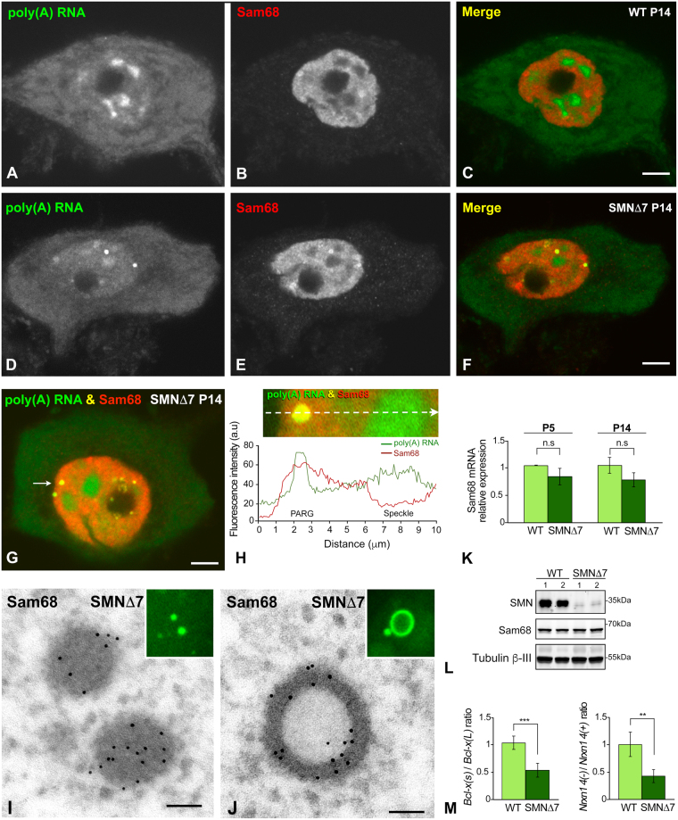
Figure 5.
(A–H) Representative examples of double labeling for poly(A) RNAs and Sam68 in WT (A–C) and SMN∆7 (D–H) MNs at P14. (A–C) In the WT MNs, Sam68 exhibits a diffuse nuclear localization with a few areas of higher intensity. (D–F) In the SMN∆7 MN, Sam68, in addition to being diffusely distributed throughout the nucleus, appears highly concentrated in two PARGs (F). Note the absence of Sam68 in nuclear speckles and the cytoplasmic depletion of poly(A) RNA. Scale bar: 3 µm. (G,H) The plot of the fluorescence intensity profiles of poly(A) RNAs and Sam68 across a line confirms the colocalization of both signals in a PARG and the concentration of poly(A) RNA, but not of Sam68, in a nuclear speckle. Scale bar: 3 µm. (I,J) Representative electron micrographs of immunogold electron microscopy localization of Sam68 in dense and ring-shaped PARGs. Scale bar: 200 nm. Insets, FISH detection of poly(A) RNAs in PARGs. (K) qRT-PCR of the relative levels of Sam68 mRNA in spinal cord extracts from WT (n = 3) and SMN∆7 mice (n = 5). No significant differences (n.s) were found when comparing WT and SMN∆7 samples during both the P5 and P14. (L) Representative western blot of Sam68 protein levels showing the dramatic reduction of SMN protein levels in spinal cord lysates from SMN∆7 mice, compared with WT mice, and the absence of significant changes in Sam68 protein levels between WT and SMN∆7 mice at the P14. (M) qRT-PCR of the Bcl-x(s)/Bcl-x(L) and Nrxn14(−)/Nrxn1 4(+) mRNA ratios in spinal cord extracts from WT and SMN∆7 mice at P14. A significant increase in the relative abundance of the Bcl-x(L) and Nrxn 4(+) splicing variants was detected in the SMN∆7 mice. p values from WT and SMN∆7 data comparison: 3.3E-4 for Bcl-x(s)/Bcl-x(L) and 4.4E-3 for Nrxn1 4(−)/Nrxn1 4(+) mRNA ratios (**p < 0.005; ***p < 0.0005).

