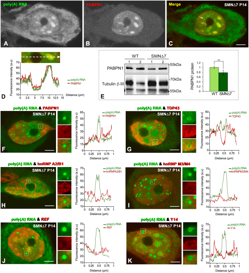
Figure 7.
(A–D) Double labeling for poly(A) RNA and PABPN1 shows the colocalization of both molecules in nuclear speckles in a PARG-free MN from an SMN∆7 mouse. Scale bar: 5 µm. (E) Representative western blotting analysis of PABPN1 levels in spinal cord lysates from WT and SMN∆7 mice. Western blot bands for PABPN1 were normalized to Tubulin ß-III, which showed double immunoreactive bands at approximately 70 kDa and 55 kDa. We choose the 55 kDa band for normalization purposes since 50–55 kDa is the predicted and apparent molecular weight of Tubulin ß-III in WB analyses. The larger 70 kDa band observed could be due to cross-reactivity with a protein related to Tubulin ß-III or a post-translationally modified form of Tubulin ß-III. The bars represent a densitometric analysis of the WB bands for PABPN1 normalized to the 55 kDa Tubulin ß-III band and expressed as the mean ± SD of three independent experiments (WT (n = 3) vs SMN∆7 (n = 5)). p value from WT and SMN∆7 data comparison: 4.3E-3 (**p < 0.005). (F–K) Double labeling for poly(A) RNAs and the RNA-binding proteins PABPN1 (F), TDP43 (G), hnRNPA2/B1 (H), hnRNPM3/M4 (I), REF (J) and Y14 (K) reveals an absence of the colocalization of poly(A) RNA with these RNA-binding proteins in PARGs, which was confirmed by plots of their respective fluorescence intensity profiles across a line. Scale bar: 4 µm.

