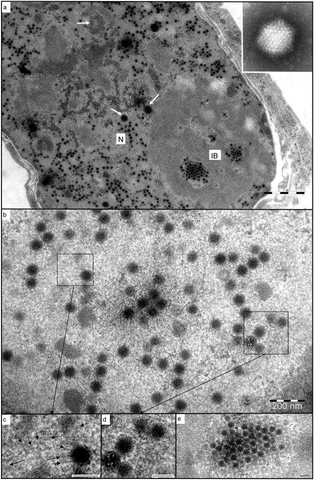
Figure 2.
Electron microscopy of RaegAdV-3085 infected Vero cells. (a) Numerous virions forming within the nucleus (N), with crystalline arrays evident within the granular matrix of an inclusion body (IB). Remnants of probable nuclear bodies are scattered throughout the nucleus (arrows). INSET: a negatively-stained virion; (b) developing virus particles within an inclusion body; (c) actin-like filaments (arrows) associated with developing virion; (d) fine, proteinaceous filaments extending between developing virions; (e) one of a series of serial sections through a crystalline array, showing the predominance of empty capsids around the periphery. Scale bars (d,e,f) = 80 nm.

