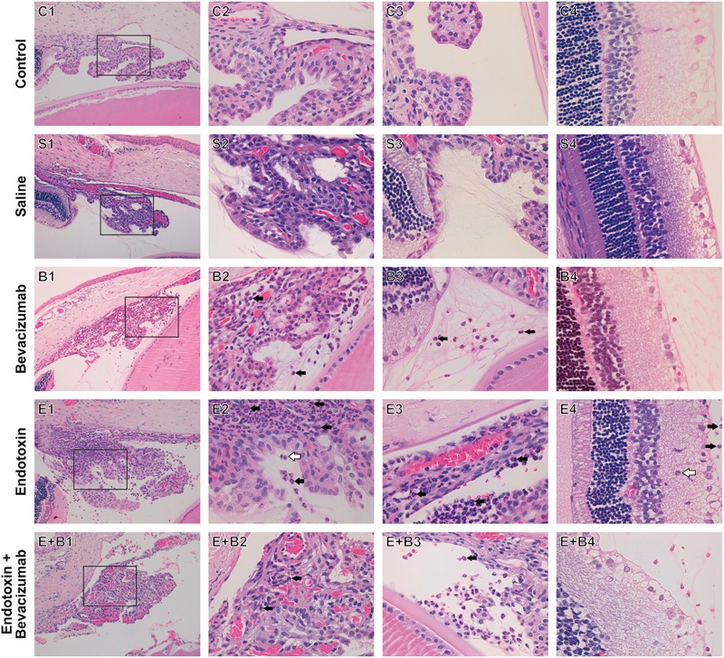
FIGURE 2.

Histopathological study of ocular structures 24 h after Bevacizumab and/or endotoxin treatment, stained with HE. Picture number 1: Ciliary body, anterior and posterior chambers, 20x. Picture number 2: Inset of picture number 1, 63x. Picture number 3: Vitreous chamber, 63x. Picture number 4: Retina, 63x. Inflammatory cells were not observed in either the Control (C) or the Saline (S) group. Inflammatory cells were seen to infiltrate extravascular uveal tissue in Bevacizumab-treated eyes (B), and reached all the studied structures, apart from the retina (ciliary body, anterior, posterior, and vitreous chambers). Many inflammatory cells neutrophils (black arrow) and monocytes/macrophages (white arrow), were found to infiltrate extravascular uveal tissue in the endotoxin-treated eyes (E) and reached all the structures under study (ciliary body, anterior, posterior and vitreous chambers, and retina). A significant reduction was noted when Bevacizumab was also injected (E+B, ciliary body, anterior, posterior and vitreous chambers, and retina).
