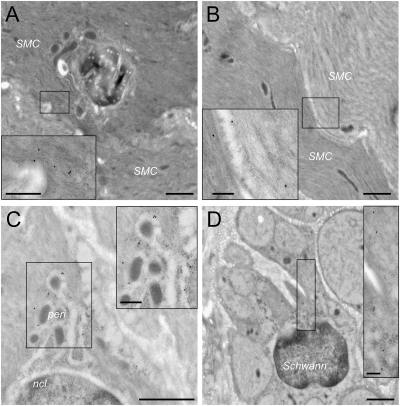
FIGURE 5.
Transmission electron microscopy confirms HCN2 presence on smooth muscle cells. (A) Typical example of HCN2 immunogold particles asymmetrically located at the border between two smooth muscle cells (SMC; magnification 7.000×, scale bar 1 μm). In the inset taken from the small box (magnification 20.000×, scale bar 250 nm), immunogold particles are indicated by white arrows. (B) Occasionally, HCN2 immunogold particles were detected symmetrically at the border between two smooth muscle cells (magnification 7.000×, scale bar 1 μm). The inset (magnification 20.000×, scale bar 250 nm) shows immunogold particles indicated by white arrows. (C) In addition to the plasma membrane compartment, HCN2 immunogold particles were also detected in the perinuclear compartment (peri) around the nucleus (ncl) containing mitochondria and rough endoplasmic reticulum (magnification 12.000, scale bar 1 μm). The inset (magnification 20.000×, scale bar 250 nm) shows immunogold particles on the endoplasmic reticulum indicated by white arrows. (D) In peripheral nerves, HCN2 immunogold particles were only detected on Schwann cells, but largely absent from the unmyelinated axons (magnification 7.000×, scale bar 1 μm). The inset (magnification 20.000×, scale bar 250 nm) shows immunogold particles within a Schwann cell cytoplasm indicated by white arrows.

