Figure 4.

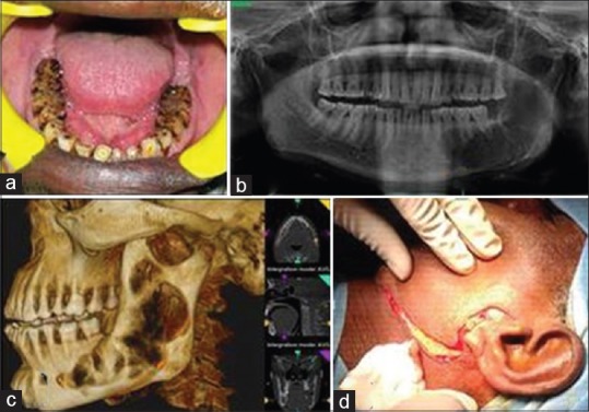
(a) Intraoral view (b) orthopantomogram (c) computed tomography with three-dimensional reconstruction view (d) preauricular, Hinds, and Risdon's incision

(a) Intraoral view (b) orthopantomogram (c) computed tomography with three-dimensional reconstruction view (d) preauricular, Hinds, and Risdon's incision