Figure 2.

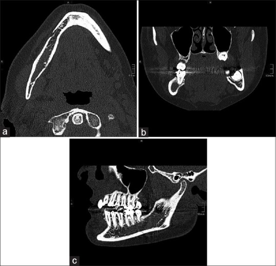
(a-c) Computed tomography: Axial, coronal, and sagittal view: Diffuse limited borders with slight erosion of the right cortical mandible and reduction of trabecular bone microstructure. The mandibular canal was breached

(a-c) Computed tomography: Axial, coronal, and sagittal view: Diffuse limited borders with slight erosion of the right cortical mandible and reduction of trabecular bone microstructure. The mandibular canal was breached