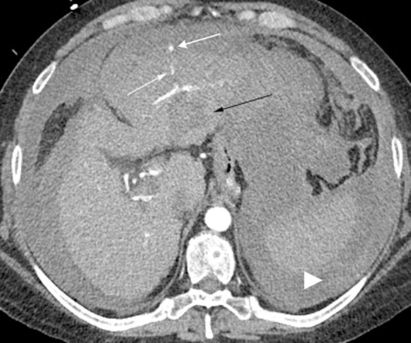
Fig. 2. Active hemorrhage.

A CT in the arterial phase shortly after HCC ablation demonstrates that active arterial extravasation (white arrows) is present, emanating anteriorly from the ablation site (black arrow). The peripheral crescent of higher attenuation (arrowhead) suggests clotted blood along with the diffuse high attenuation peritoneal blood.
