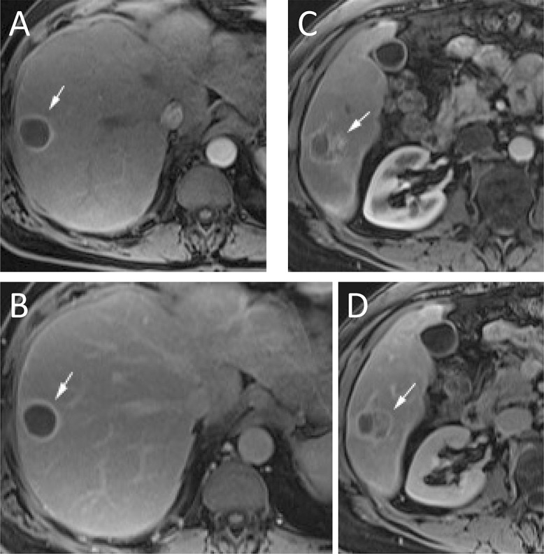
Fig. 4. PostTACE treatment.

No residual tumor (A and B) and residual disease present (C and D). (A) Arterial phase imaging, 1 month after TACE demonstrates a thin (<5 mm) rim (arrow) of enhancement without nodularity. (B) The thin rim of enhancement (arrow) persists on portal venous phase imaging. These findings are consistent with expected post treatment changes and not recurrence. (C) In another patient, arterial phase imaging, 1 month after TACE demonstrates a nodular (>5 mm) area of enhancement (arrow). (D) This nodular area of enhancement (arrow) demonstrates washout on portal venous phase imaging. These findings are consistent with recurrent HCC. Abbreviations: HCC, hepatocellular carcinoma; TACE, transarterial chemoembolization.
