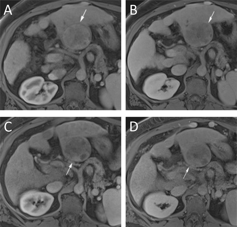
Fig. 5. Transarterial radioembolization treatment imaging findings.

One month imaging shows diffuse patchy enhancement on arterial (A) and, to a lesser extent, on portal venous (B) phase imaging (arrows). While multiple areas of patchy enhancement are present at 1 month, the 3 months imaging shows an area of enhancement on arterial (C) and washout on portal venous (D) imaging (arrows). Because of these MRI findings, this lesion was considered residual disease and treated again without histologic confirmation.
