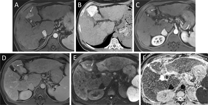
Fig. 7. DWI to help diagnosis post TACE residual.
(A) Prior to treatment, an early arterial phase MRI demonstrates a hyperenhancing lobulated mass (arrow) in segment 4. Delayed phase washout and DWI sequences (not shown) established the imaging diagnosis of HCC. (B) Six weeks later this patient underwent conventional TACE. The CT following this procedure shows complete saturation of the HCC area with lipiodol. (C) Late arterial phase MRI one month later shows a continued hyperenhanced area (arrow). (D) A 3-minute delay image demonstrates washout (arrow). (E, F) The DWI series with correlating b 600 (E) and ADC (F) sequences confirm this area as residual HCC with high SI and low SI respectively (arrows). “g” marks the gallbladder. Abbreviations: ADC, apparent diffusion coefficient; CT, computed tomography; DWI, diffusion weighted imaging; HCC, hepatocellular carcinoma; MRI, magnetic resonance imaging.

