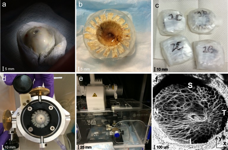
Figure 1.
Experimental setup for the inflation test of the human LC showing the (a) ONH cut flush to the sclera; (b) ring-mounted posterior scleral cup; (c) posterior scleral samples incubating at 37°C; (d) custom inflation holder; (e) Zeiss LSM 710; and (f) maximum intensity projection, SHG image of the LC.

