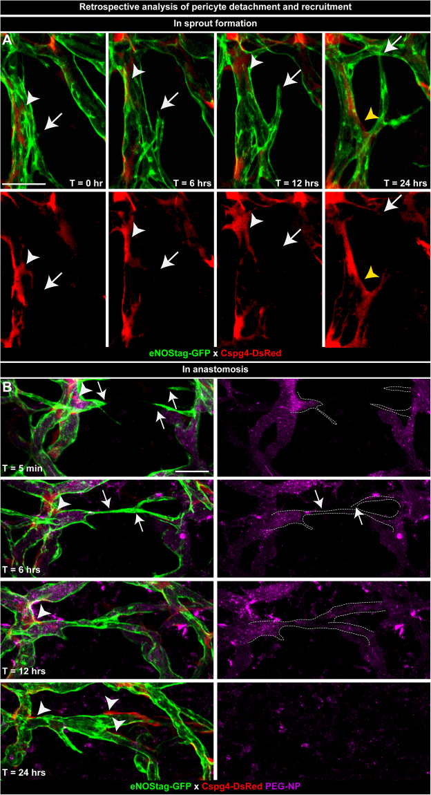
Figure 5.
Retrospective analysis of pericyte detachment and recruitment. Images of vessel development taken at T = 0, 6, 12 and 24 hrs were re-evaluated. (A) Subsequential projection of a developing endothelial sprout (arrow) and an adjacent dispersing (white arrowhead) and re-emerging (yellow arrowhead) pericyte. (B) Forward movement of endothelial cells (arrow) and recruitment of pericytes (arrowhead) during anastomosis. Blood flow is detected using pegylated nanoparticles (PEG-NP) that remain in the blood circulation for approximately 20 hrs. Scale bars represent 50 µm.

