Figure 6.
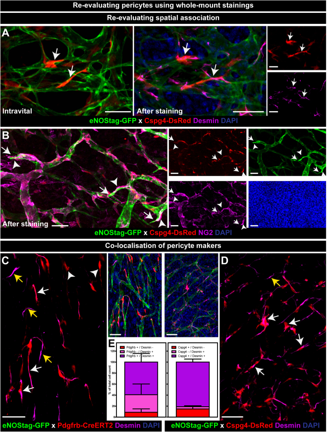
Re-evaluating pericytes using whole-mount staining. (A,B) Re-evaluating spatial endothelial cell – pericyte association. (A) Intravital images of Cspg4+ pericytes shown in Fig. 4 re-evaluated after desmin staining (arrow). (B) Cspg4+ pericytes re-stained with NG2 (arrowheads) shown in close proximity of endothelial tip cells (arrow). (C–E) Co-localization of pericyte markers. (C,D) Whole-mount staining of tumor tissue showed Pdgfrb+ or Cspg4+ pericytes co-expressing desmin (white arrow), Pdgfrb+ pericytes not stained for desmin (arrowhead), and Pdgfrb- or Cspg4- pericytes stained positive for desmin (yellow arrow). (E) Quantification of co-expression of Pdgfrb or Cspg4 with desmin. Data represent mean ± SEM. Scale bars represent 50 µm.

