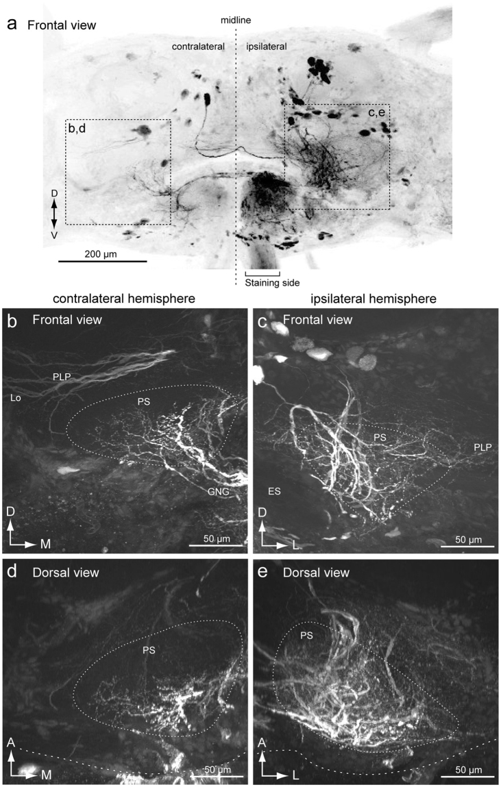
Figure 3.
Mass-staining result by backfilling from one side of the neck connective. (a) Maximum intensity projection of mass-staining result. The frontal view is shown. The side of filling is noted. (b,c) Frontal view of the innervation in the posterior slope (PS) of the contralateral (b) and ipsilateral sides (c). Wide field innervation is observed in the ipsilateral hemisphere. (d,e) Dorsal view of the innervation in the PS of the contralateral (d) and ipsilateral sides (e). The innervation is extended more anteriorly in the ipsilateral hemisphere. ES, esophagus; GNG, gnathal ganglia; PLP, posterior lateral protocerebrum.

