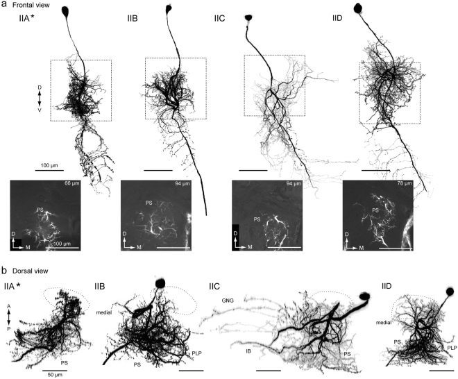
Figure 7.
Group-II DNs innervate the PS. (a) Morphology of group-II DNs. Maximum intensity projection of all innervation in the brain (top) and confocal stack of the neurite innervation in the posterior slope (PS) are shown (bottom). The frontal view is shown. A flipped image is shown for comparison (asterisk). All DN types have innervation in the medial PS. (b) Drosal view of DN innervation in the brain. The LAL is shown with a broken line. The innervated areas are similar among group-II DNs, in comparison with group-I DNs (Fig. 4). GNG, gnathal ganglion; IB, inferior bridge; PLP, posterior lateral protocerebrum; PS, posterior slope.

