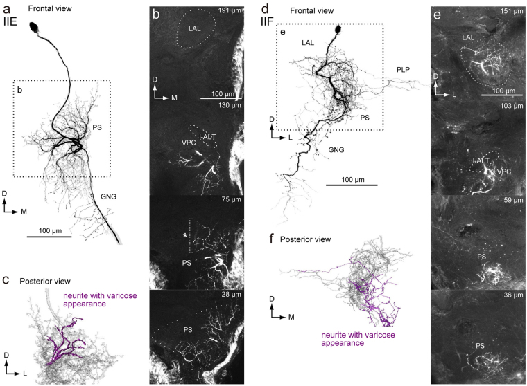
Figure 8.
Newly identified group-II DNs. (a) Maximum intensity projection of the DN innervation in the brain. The morphology is similar with the group-IID DN. Unlike to other group-II DNs, the DN has innervation with varicose appearance in the PS. The DN also innervates the PLP. (b) Confocal stacks for the DNs in (a). The DN has innervation in the almost all of the LAL, the ventral protocerebrum (VPC) and the medial part of the inferior PS. The depth from the posterior surface is shown in the top-right. (c) Posterior view of three-dimensional reconstruction of the DN in the PS. The neurites with varicose appearance are shown in magenta. (d) Maximum intensity projection of the DN innervation in the brain. The morphology is with the group-IIC DN. Unlike other group-II DNs, the DN has innervation with varicose appearance in the PS. (e) Confocal stacks for the DNs in (a). The DN has innervation in the almost all of the LAL, the VPC and the inferior PS. The depth from the posterior surface is shown in the top-right. (f) Posterior view of three-dimensional reconstruction of the DN in the PS. The neurites with varicose appearance are shown in magenta.

