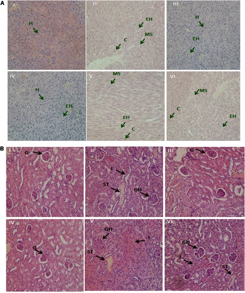
FIGURE 5.

(A) The demonstrative micrographs of the hepatic (liver) tissues of the mice with hematoxylin-eosin (H&E staining; magnifications × 400). (I) Control, (II) Lead only, (III) Non-encapsulated L. plantarum KLDS 1.0344 only, (IV) PRS encapsulated L. plantarum KLDS 1.0344 only, (V) Non-encapsulated L. plantarum KLDS 1.0344 plus lead, (VI) PRS encapsulated L. plantarum KLDS 1.0344 plus lead. Exposure to lead produced histological alterations in the liver, comprising chromatin condensation (C), injury of intact liver plates and cytoplasmic vacuolization. While in case of liver histopathological examination the lead only group (II), showed vacuolar degeneration, enlarged hepatocytes (EH) and microvesicular steatosis (MS). No obvious histopathological changes were noticed in control (I). While with the exemption of certain hepatocytes (H) swelled observed co-treatment with Non-encapsulated and PRS encapsulated KLDS 1.0344 with lead indicated protecting properties against identical liver injuries (V, VI). (B) Demonstrative micrographs of renal (kidney) tissues of the mice with hematoxylin-eosin (H&E staining; magnifications × 400). (I) Control, (II) Lead only, (III) Non-encapsulated L. plantarum KLDS 1.0344 only, (IV) PRS encapsulated L. plantarum KLDS 1.0344 only, (V) Non-encapsulated L. plantarum KLDS 1.0344 plus lead, (VI) PRS encapsulated L. plantarum KLDS 1.0344 plus lead. There was normal histomorphology of the kidney was obviously apparent in the control groups (I). Treatments with Non-encapsulated and PRS-based encapsulated KLDS 1.0344 with lead considerably relieved such renal damages. While in lead only group (II) the volume of the glomeruli (G) turned significantly bigger i.e. glomeruli were hyperemic, while some of the glomeruli were found missing, swelling was also observed in some renal tubular epithelial cells (II, IV,VI). Symptoms like glomerular hyperemic (GH), granular degeneration, swollen tubular (ST) epithelial cells and inflammatory cells (I) of all treated groups (III, IV, V, VI) were improved as compared to the lead (Pb) group.
