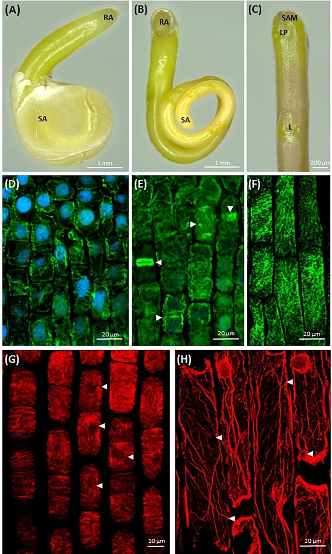
FIGURE 1.
Morphology of dodder seedlings and maximum intensity projection images of cytoskeleton arrays in shoot apex cells: (A,B) bipolar filiform seedling of C. monogyna devoid of cotyledons, spirally coiled with a tapered shoot apex (SA) covered with testa’s remnants or released, and a blunt radicular end (RA), 2nd day post-germination; (C) shoot apex of C. monogyna with scale-like leaf (L), leaf primordia (LP), and shoot apical meristem (SAM); 4th day; (D) cortical and endoplasmic microtubules (green) in C. europaea shoot apical meristem cells, DAPI-stained nuclei (blue), 7th day; (E) endoplasmic microtubules, mitotic spindles, phragmoplast and two daughter cells after cytokinesis (arrowheads) in C. europaea shoot apical meristem, DAPI-stained nuclei, 7th day; (F) cortical microtubules in C. monogyna shoot cortical cells; (G) actin filaments in C. europaea shoot apical meristem cells, 7th day; (H) actin filaments in C. monogyna shoot cortical cells, 7th day.

