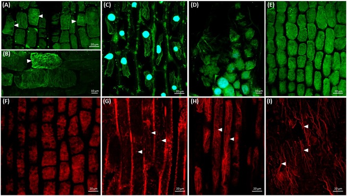
FIGURE 5.
Arrays of cortical microtubules (green) and actin filaments (red) in Cuscuta cells. Nuclei (blue) are stained with DAPI. (A,B) Taxol (10 μM) stabilized cortical microtubules (arrowheads) in shoot cells; (C) taxol (10 μM) stabilized microtubules in root-like structure cells, 7th day; (D) oryzalin (10 μM) induced disruption of microtubules in shoot apical meristem cells; (E) oryzalin (10 μM) induced disruption of microtubules in shoot cortical cells; (F) cytochalasin B (100 μM) induced disruption of actin filaments in shoot apical meristem cells; (G) cytochalasin B (100 μM) induced fragmentation of actin filaments in shoot cortical cells; (H) longitudinally oriented F-actin cables and cortical transverse actin arrays in shoot cortical cells after BDM (20 mM) treatment; (I) disturbed organization of F-actin cables in root-like structure plasmolyzed cells after BDM (20 mM) treatment, 7th day.

