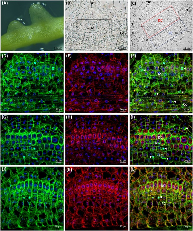
FIGURE 8.
Development of C. europaea prehaustorium initiated by a first contact with Nicotiana benthamiana as a susceptible host: (A) general morphology; (B) cross-section of prehaustorial bulge: meristematic centre (MC) and the cortex (Co) cells; (C) significantly extended distal digitate cells (DC) and the proximal compact file cells (FC). The sites of contact with the host are marked by asterisks (∗). Median longitudinal sections of C. europaea prehaustorium depicting immunolabeled arrays of microtubules (D,G,J, green), actin filaments (E,H,K, red), and their combination (F,I,L, merge). Nuclei are stained blue with DAPI. Dividing cells are marked by arrowheads ( ): (D–F) meristematic centre (MC) of prehaustorium located close to xylem vessels in cortex (Co) cells, early developmental stages; (G–I) file (FC) and digitate (DC) cells composing the essential part of prehaustorium; (J–L) significantly prolonged DC and still dividing FC are present in late developmental stages.

