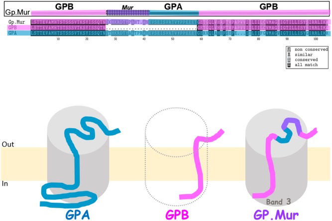
FIGURE 1.
Glycophorin B-A-B hybrid protein GP.Mur interacts with band 3. (Top) Protein sequence alignment of GPA, GPB, and GP.Mur, which was first published in Blood (Hsu et al., 2009), is modified here with color coding. The sequences of GPA and GPB are color-coded with blue and pink, respectively. The antigenic Mur peptide at the cross-over region in GP.Mur is color-coded purple. The configuration of glycophorin B-A-B is revealed by the combination of pink-(purple)-blue-pink colors. (Bottom) GPA, GPB, and GP.Mur are homologous membrane proteins, each with a single transmembrane span. Their N-terminal sequences are located extracellular, with heavy glycosylation (not shown); their C-terminal sequences are intracellular. Both GPA and GP.Mur interact with band 3 (gray cylinder), and GPB does not.

