Figure 3.

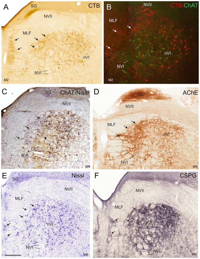
Detailed views of corresponding planes of the right nVI in transverse monkey sections from different experiments. (A,B) Identification of INTs via retrograde labeling with choleratoxin subunit B (CTB) injection into the oculomotor nucleus (nIII; A,B, red) and their absence of ChAT-immunoreactivity (B, green). Anterograde CTB-labeling from nIII highlights (A) the paramedian tractneurons (PMT) in the supragenual nucleus (SG) and (B) the “intrafascicular nucleus of the preabducens area” forming bridges between the medial nVI and midline (A–F; arrows). The PMT neurons are not ChAT-positive (C, arrows), but are highlighted by acetylcholinesterase staining (AChE) in the SG and the “intrafascicular nucleus of the preabducens area” (D, arrows). The appearance of PMT neurons is further demonstrated in corresponding sections stained for Nissl (E, arrows) and CSPG (F, arrows). Scale bar = 500 μm in (E; applies to A–F). MLF, medial longitudinal fascicle; NVI, abducens nerve.
