. 2016 May 11;7(8):4990–5001. doi: 10.1039/c6sc00696e

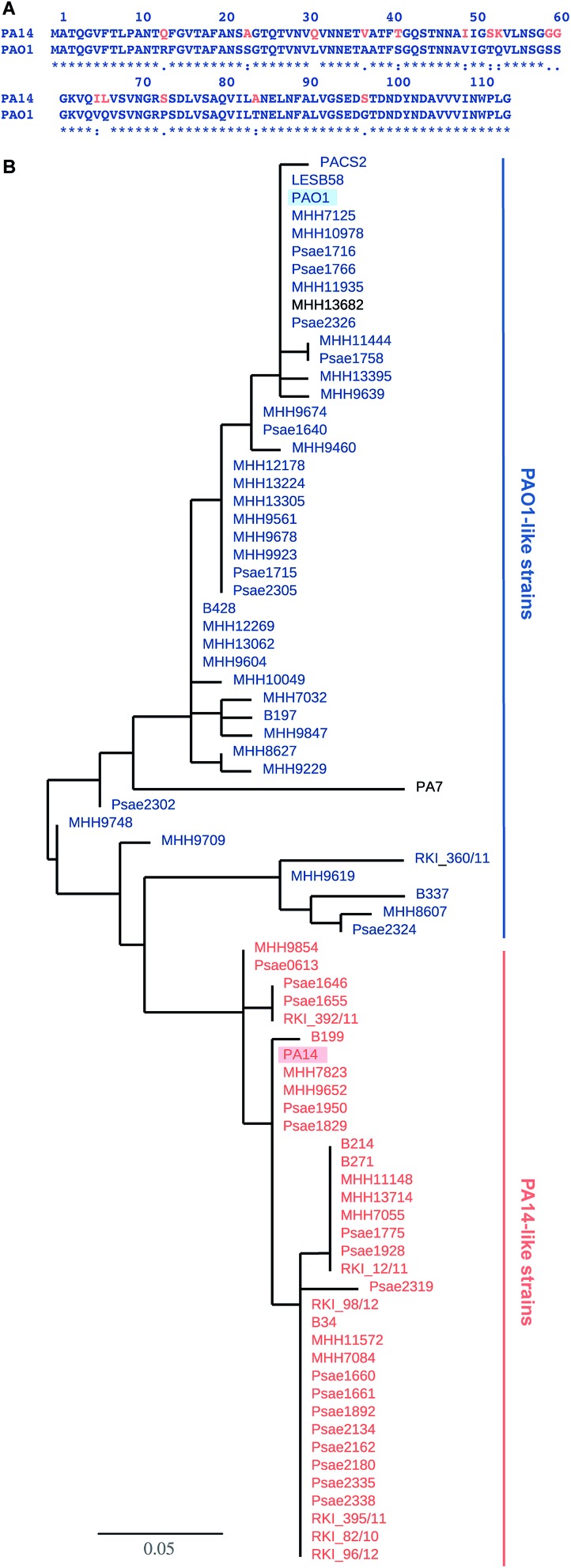
Fig. 1. LecB sequence comparison: (A) alignment of LecB sequences from PA14 and PAO1. Amino acid numbering corresponds to the crystal structure of LecBPAO1, which lacks methionine; (B) phylogenetic tree based on LecB sequences from P. aeruginosa clinical isolates and reference strains PAO1, PA7, PA14, PACS2, LESB58. An independent phylogenetic analysis based on 200 core gene sequences allowed the classification of strains as PA14- or PAO1-like strains, illustrated in the color coding in orange (PA14) and blue (PAO1), PA7 and MHH13682 (shown in black) could not be assigned to either strain family. The branch length is proportional to the number of substitutions per site.

Fig. 1