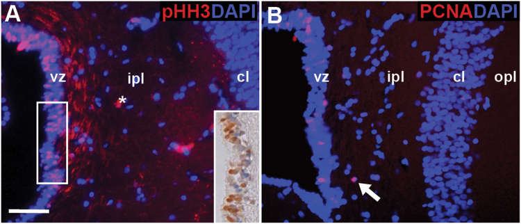
Figure 4.
Ventricular zone cells express proliferation markers phosphorylated histone -H3 (pHH3) and proliferating cell nuclear antigen (PCNA). (A) Within the sulcus septomedialis, pHH3+ cells are restricted to the ventricular zone. Expression was confirmed using immunohistochemistry (inset). (B) Likewise, PCNA+ cells are most abundant in the ventricular zone, although occasionally observed in close contact to the ventricular zone within the inner plexiform layer (white arrow). Neither pHH3+ nor PCNA+ cells were ever observed within the cellular layer. Asterisk indicates an artifact. Scale bar A: 10 μm. cl = cellular layer, ipl = inner plexiform layer, opl = outer plexiform layer, vz = ventricular zone.

