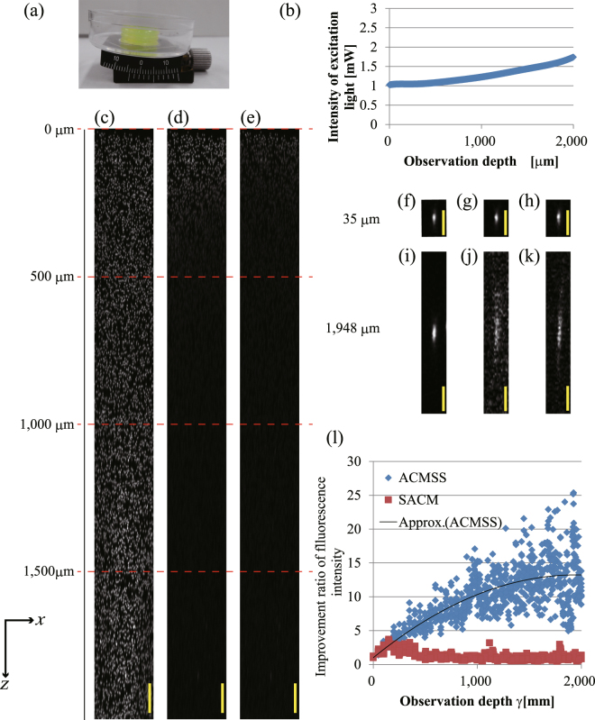
Figure 2.
Observation results of fluorescent beads of 3-μm diameter in a transparent epoxy resin tilted at 0.0873 rad (5°). (a) Photograph of the transparent epoxy resin tilted by a goniometric rotation stage. (b) Excitation light intensity. The excitation light intensity was measured under the objective lens. (c–e) -projected images for an optical depth of 0 μm to 2,000 μm obtained from scans performed with wavefront modulation using ACMSS, with wavefront modulation using SACM, and without wavefront modulation, respectively. (f–h) image of the observed bead at a 35-μm optical depth when a TPM scan was performed with wavefront modulation using ACMSS, with wavefront modulation using SACM, and without wavefront modulation, respectively. (i–k) image of the observed bead at a 1,948-μm optical depth. (l) Quantitative evaluation of the improvement in the fluorescence intensity from the fluorescent beads. The scale bars indicate 100 μm in (c–e) and 20 μm in(f–k).

