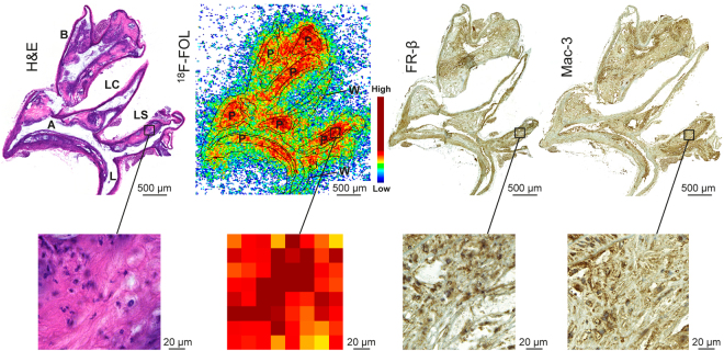
Figure 4.
Ex vivo 18F-FOL autoradiography, histology and immunohistochemistry of mouse aorta. Representative hematoxylin-eosin (H&E), 18F-FOL ex vivo autoradiograph, FR-β and Mac-3 immunohistochemical staining (macrophages) of adjacent aorta cryosections. The images on the bottom show high-power views of the area within the black rectangle on the upper images. A = arch; B = brachiocephalic artery; L = lumen; LC = left common carotid artery; LS = left subclavian artery. P = plaque; W = healthy vessel wall.

