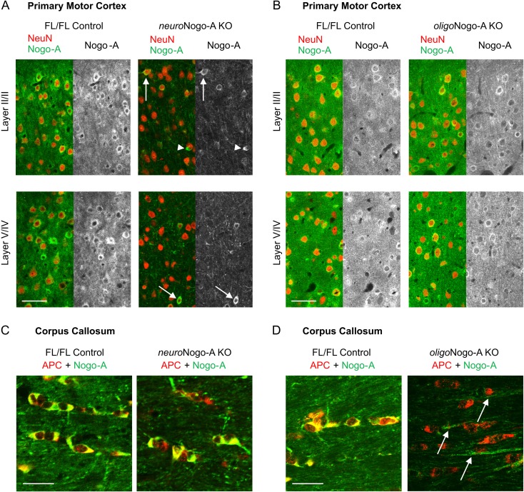
Figure 1.
Nogo-A expression in motor cortex (M1) and CC of neuroNogo-A KO and oligoNogo-A KO mice. (A) Absence of Nogo-A-immunostaining in the majority of neurons (due to thy-1-Cre expression) in neuroNogo-A KO mice. Nogo-A expression persists in the oligodendrocytes (arrowheads) and in a small population of NeuN positive neurons (arrows),probably due to their lack of thy-1-Cre expression. (B) Neuronal Nogo-A is preserved in M1 cells of oligoNogo-A KO mice. (C) In the CC, APC-positive oligodendrocytes express Nogo-A in control and neuroNogo-A KO mice. (D) APC-positive oligodendrocytes lack Nogo-A in the oligoNogo-A KO mice. The arrows indicate Nogo-A positive axons. Calibration bar: 50 μm in (A, B); 25 μm in (C, D).

