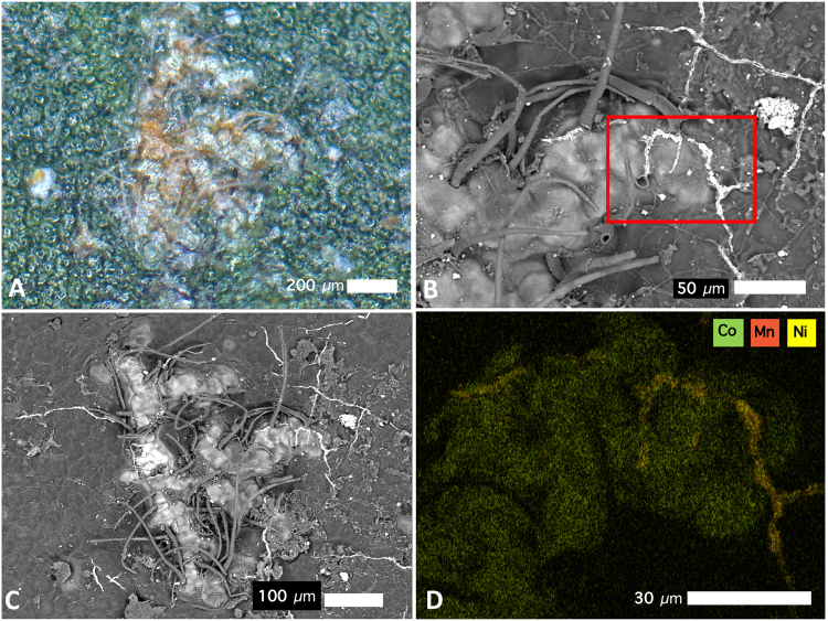
Figure 2.
Cobalt-rich surface lesions on the abaxial leaf surface of Glochidion cf. sericeum: (A) bright-field light microscopy image showing brown lesions developed under the cuticle; (B) Scanning Electron Microscopy (SEM) image showing the same lesion with trichomes; (C) SEM image of the same lesion, full view; (D) Energy-Dispersive Spectroscopy (EDS) image of the lesion with rectangular box outline field-of-view in relation to the SEM image in b. Key to colours: Green is Co, orange is Mn, yellow is Ni. Scale bars denote 30, 50, 100 and 200 μm.

