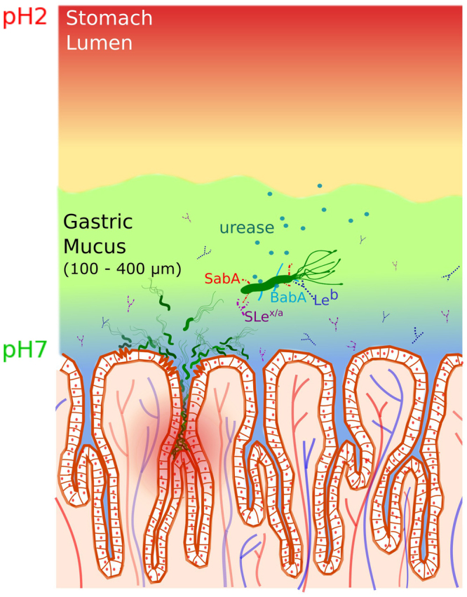
Figure 1.

A schematic illustration of the gastric mucosa depicting the interaction of H. pylori with mucin. The cells on the mucosal epithelial surface are shown demarcating the crypts and the underlying vasculature. The pH gradient from 7 at the cell surface to about 2 in the lumen is indicated by the blue to red shading. A single bacterium is greatly enlarged to display the adhesins, BabA (blue) and SabA (red) which bind to specific antigens Leb (blue symbol) and SLex/a (purple symbol) on the mucin, respectively. The urease (green circles) secreted by the bacterium enables it to hydrolyze acid, de-gel the mucus and swim across (see text). Bacteria colonizing on the cell surface as well as deep in the crypts are also shown.
