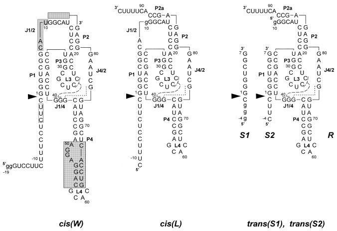
Figure 1.
Secondary structure models of cis- and trans-acting antigenomic delta ribozymes. Numbering of nucleotides corresponds to the wild-type ribozyme sequence and nucleotide changes are shown in lower case letters. Base paired segments are denoted P1–P4 and single-stranded regions as J1/2, J1/4 and J4/2. Nucleotides connected with broken and dotted lines indicate the 2 bp helix P1.1 and non-standard G-G interactions analogous to those found in the crystal structure of the genomic variant. Catalytic cleavage sites are marked by filled triangles. The shaded segments in cis(W) denote regions changed in the ribozyme variants.

