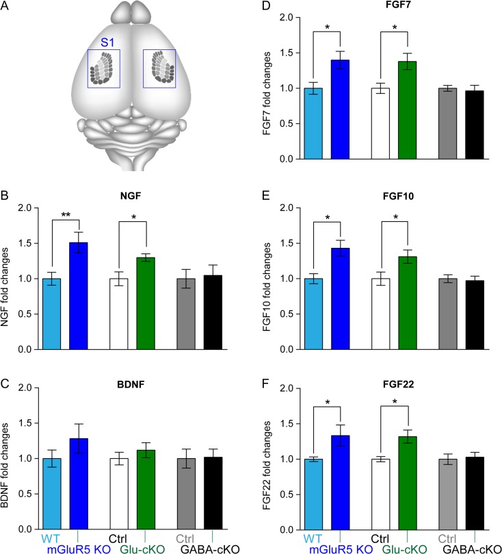
Figure 1.
mGluR5 deletion in glutamatergic neurons results in increased mRNA levels of Ngf, Fgf7/10/22, but not Bdnf. (A) Schematic illustration shows the location of whisker-barrels (individual rows are labeled with different shades of gray) in the brain. Blue boxes indicate the cortical area collected for qPCR and Western analysis. Bar graphs show the normalized levels of Ngf (B), Bdnf (C), Fgf7 (D), Fgf10 (E), and Fgf22 (F) mRNA (n ≥ 5 pairs per transgenic mouse line). Mann–Whitney test was used to assess statistical significance. The statistical analysis (*) refers to wild-type or control group compared to mGluR5 global KO, Glu-mGluR5-cKO (NEX-Cre positive), or GABA-GluR5-cKO (DLX-Cre positive) group. *P < 0.05.

