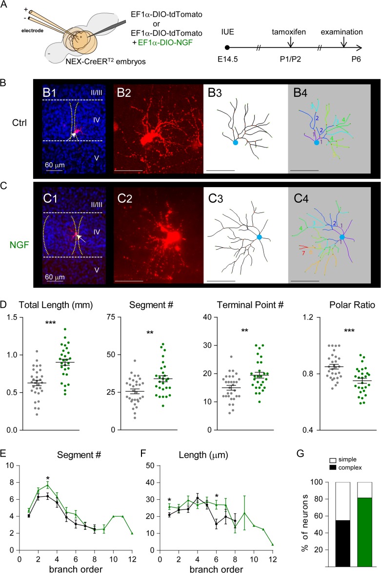
Figure 4.
Postnatal NGF expression influences dendritic patterning of layer IV cortical neurons. (A) Schematic representation of the electroporation and tamoxifen treatment protocols that were used to express NGF. (B) Examples of original images and computer-aided reconstructions. B1 and C1 show the locations of barrels (dashed lines) and reconstructed neurons (white arrows). II–V, indicate cortical layers. The projected images from confocal image stacks are shown in B2 and C2. B3 and C3 show the traced images of neurons in B2 and C2. B4 and C4 show color-coded segments according to their branch orders. (D) The total length, segment number, and terminal point of dendrites were all significantly increased in NGF-overexpressing neurons. The dendritic polarity ratio was significantly lower in NGF-overexpressing neurons. Summaries for segment number (E) and length (F) per branch order. (G) Summary of neuron complexity determined by grouping branch order <5 (simple neurons) or branch order ≥5 (complex neurons). Statistical analysis: Mann–Whitney test. The statistical analysis (*) compared NGF-expressing group to control (Ctrl) group. *P < 0.05; **P < 0.01; ***P < 0.001.

