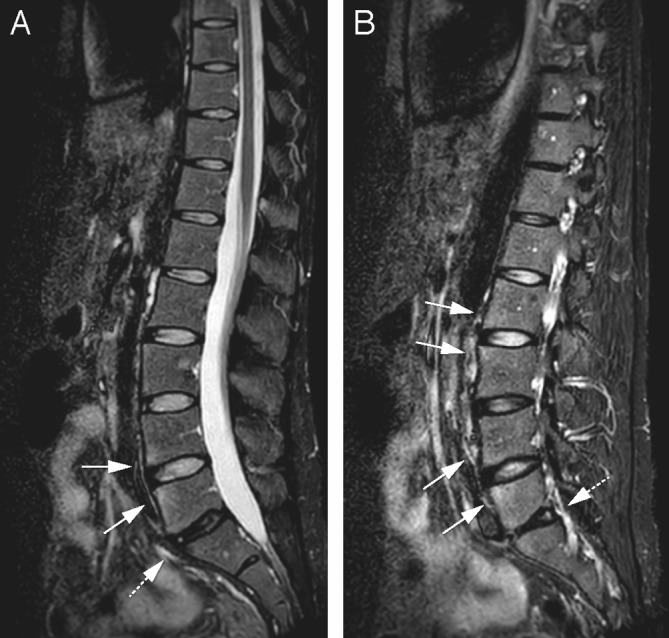
Figure 3.

Same patient as 1b. On another sagittal short tau inversion recovery slice, the high signal intensity at the corners of the endplates, signs of axial spondyloarthritis, are more clearly seen at L2–3 as well (arrows). The degenerated disc L5–S1 (dotted arrow) has a high-intensity zone at the posterior part (B).
