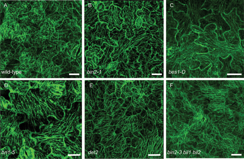
Fig. 2.
BR mutants showed parallel arrangement of microtubules. (A–F) Cortical microtubules of cotyledon pavement cells in wild-type (A), bin2-1 (B), bes1-D (C), bri1-5 (D), det2 (E), and bin2-3 bil1 bil2 triple mutant (F) with a transgene expressing GFP–tubulin in the background for all lines. Bars: 20 μm. (This figure is available in color at JXB online.)

