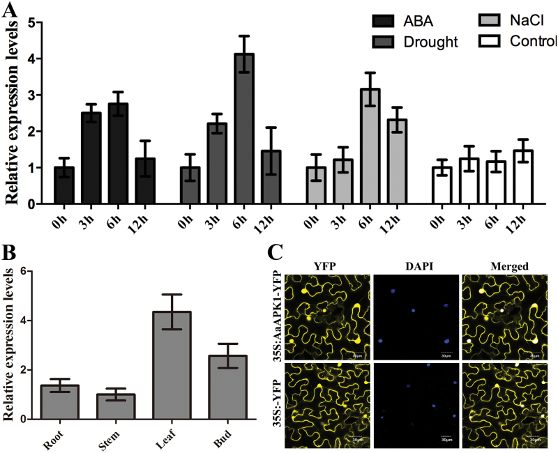
Fig. 3.
Expression analysis and subcellular localization of AaAPK1. (A) The expression levels of AaAPK1 in under treatment with ABA, drought (air-drying), and NaCl. (B) The tissue profiles of AaAPK1. (C) Subcellular localization of AaAPK1. The coding sequence of AaAPK1 was in-frame fused with YFP, and under the control of the 35S promoter, then transferred into A. tumefaciens GV3101 and infiltrated into leaves of N. benthamiana. DAPI-stained nuclei were used as the control. (This figure is available in colour at JXB online.)

