Figure 6.
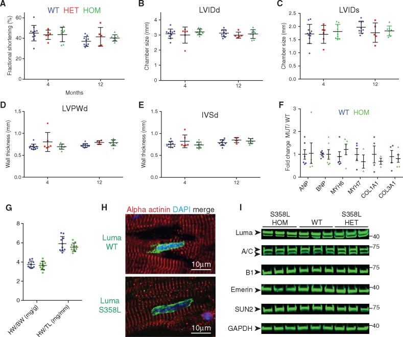
Luma S358L KI mice display no overt defects in cardiac function and morphology. Echocardiographic measurements of (A) FS, (B) LVIDd, (C) LVIDs, (D) LVPWd, and (E) IVSd over the course of 12 months in WT (n = 11; 9 females, 2 males), Luma S358L heterozygous (HET) mice (n = 6; 3 females, 3 males), Luma S358L homozygous (HOM) (n = 9; 5 females, 4 males). (F) Quantitative real-time PCR analysis of foetal genes, ANP, BNP, Myh6, Myh7, and pro-fibrotic markers, Collagen 1a1 and 3a1, in WT and Luma S358L homozygous mouse hearts at 2–3 months (n = 3–6 per genotype). (G) HW/BW and HW/TL ratios of WT vs. Luma KO mice at 47 weeks (n = 10 per genotype). (H) Immunofluorescence analysis of adult hearts isolated from WT and S358L homozygous mice. Sections were stained with antibodies raised against Luma (green), alpha actinin (red), and DAPI (blue). Note that Luma S358L has a similar localization to WT Luma. (I) Western blot analysis of Lamins A/C (A/C), B1 (B1), Emerin, SUN2, and Luma in WT and Luma S358L KI hearts. Glyceraldehyde 3-phosphate dehydrogenase (GAPDH) serves as a loading control. Arrowheads show the predicted MW of the indicated proteins. MW is indicated in kDa (n = 3 per genotype). P > 0.05 according to two-way ANOVA with Sidak’s post-hoc test (A–G).

