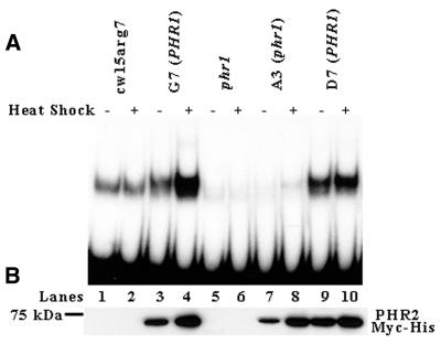
Figure 5.
PHR2 Myc-His overexpressed in a phr1 background is largely inactive. (A) Equivalent amounts of protein from ammonium sulfate extracts of heat shocked and non-heat shocked cw15arg7, G7 (PHR1), phr1, A3 (phr1) and D7 (PHR1) cells were incubated in the dark at 25°C for 30 min with radioactively labeled DNA probe that had been UV irradiated. The mixtures were then run on a 5% non-denaturing polyacrylamide gel and visualized by autoradiography. (B) Western blot analysis of ammonium sulfate protein extracts from cw15arg7, G7 (PHR1), phr1, A3 (phr1) and D7 (PHR1) cells before and after heat shock. Equal amounts of protein was separated by SDS–PAGE, transferred to PVDF and blotted with anti-Myc antibody. The position of the 75 kDa marker is shown on the left.

