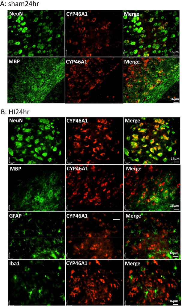
Figure 3.
Expression of CYP46A1 in the neurons and oligodendrocytes. Images of double Immunofluorescent staining with CYP46A1 antibody (red, middle panels) paired with another antibody specific for neuron (NeuN), oligodendrocyte (MBP), astrocyte (GFAP), or microglia (Iba1), shown in green on the left panels. The representative images from the sham (A) and HI-injured animals at 24hr after HI (B).

