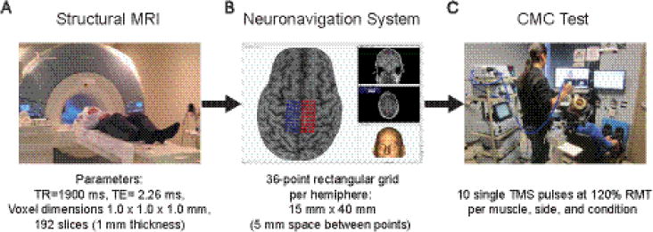
Figure 1.

Flowchart of the experimental procedures prior to first session (A-B) and during both experimental sessions (C).
A) In a Siemens 3T TIM trio scanner (Siemens, Erlanger, Germany), participants were asked to keep still and wore earplugs to attenuate the scanner’s loud noise. B) Using Brainsight™ TMS frameless stereotaxy neuronavigation system, each participant’s MRI was co-registered to the anterior and posterior commissures, so each MRI could be mapped using the Montreal Neurological Institute atlas. Three-dimensional skin and curvilinear brain models were reconstructed. A skin model was used to identify the tip of the nose, nasion, and supratragic notch of the right and left ear, which were used to calculate the participant to image registration. A curvilinear brain model was used to manually identify the leg motor area on which a rectangular grid was overlaid below the cortical surface on each hemisphere. C) Single pulse TMS was applied bilaterally over the SOL and TA optimal hotspot using Brainsight™ while muscle responses were collected simultaneously from both legs during rest and active conditions. Both feet were secured in boots (7.5° of plantarflexion) to ensure a consistent lower extremity posture across participants and sessions, as well as ease of isometric ankle contractions. Additionally, to minimize any hip or knee motions, participants’ thighs were firmly strapped to the leg support pads.
