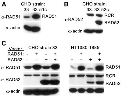
Figure 2.
Western blot analysis of overexpressed human RAD51 and RAD52. For detection of RAD51 and RAD52, we loaded 20 and 60 µg, respectively, of protein from whole cell extracts. Proteins were separated by SDS–PAGE, transferred to PVDF membranes and detected with polyclonal antibodies to RAD51 (α-RAD51) or RAD52 (α-RAD52) using ECL reagents (Amersham Pharmacia Biotech, Piscataway, NJ). Parallel lanes were probed with anti-β-actin antibodies as loading controls. (A) Endogenous RAD51 levels in CHO strain 33 (left lane) and the RAD51 constitutive overexpression strain 33-51c (right lane). (B) Endogenous RAD52 in strain 33 is too low to be detected in this exposure (left lane), but easily detected in the RAD52 constitutive overexpression strain 33-52c (right lane). An α-RAD52 cross-reacting (RCR) species of higher molecular weight was also detected. (C) RAD51 and/or RAD52 expression vectors were transfected into strain 33 or HT1080-1885 as indicated and cell extracts were prepared 48 h post-transfection for western blot analysis as above.

