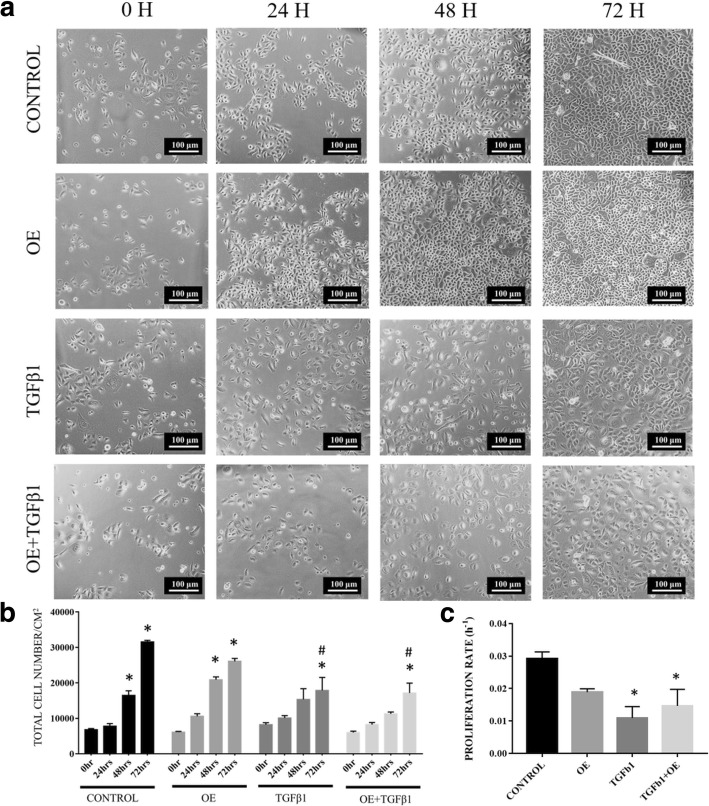
Fig. 4.
a Distribution of untreated RECs (control), 0.05% (v/v) OE-treated RECs (OE), RECs induced to EMT with 5 ng/ml TGFβ1 (TGFβ1) and RECs induced to EMT with TGFβ1 and treated with OE (TGFβ1 + OE) from 0 h to 72 h. b Total cell number/cm2 versus (vs) time per group. *P < 0.05 indicates significant difference when compared with 0 h and # P < 0.05 indicates significant difference when compared with the control group. c The proliferation rate of cells. *P < 0.05 indicates significant difference when compared with the control

